Slideshow
Art in Research winning images announced

On June 7, the Yale Postdoctoral Association and the Office of the Provost announced the winners of the inaugural Art in Research Competition.
Winners were selected in a variety of categories, including research microphotography, research macrophotography, and research illustrations, as well as images related to top Yale science priorities: data science, neuroscience, inflammation, and environmental and climate science.
The competition was open to postdoctoral researchers, associate research scientists, and graduate students. The organizers said the contest drew more than 60 submissions.
Winners received gift cards and their winning images may be used to showcase the diversity of research at Yale in future university announcements and promotional materials, and by external media outlets.
Browse the slideshow at top to view the winning images and read their full descriptions below.
Winners
“Actin network”
Camelia Muresan, from the Department of Biomedical Engineering and the lab of Michael Murrell, won first place in the micro art category and first place in the inflammation category. The image was acquired with a fluorescent microscope and it shows the structure of the F-actin network. In living systems actin regulates fundamental processes such as cell division, migration, membrane protrusions, intracellular interactions, and force generation. Alterations of the actin cytoskeleton can result in autoinflammatory disease.
“Zinc oxide salt microparticles imaged using Scanning Electron Microsopy”
Rita Matta, from the Department of Biomedical Engineering and the lab of Anjelica Gonzalez, won second place in the micro art category. Better understanding of mechanisms that promote neural stem cell recruitment and differentiation are important factors to create successful stem cell therapy. Understanding the signaling clues and soluble factors that promote neural stem cell migration can provide insight for tissue engineers and neurologists to create a therapy which can enhance cellular response to brain tissue damage.
“Cell escape from tumor”
Muhammad Yousafzai, from the Department of Biomedical Engineering and the lab of Michael Murrell, won third place in the micro art category. The onset of migration from a 3D tumor is imaged with a confocal microscope. The actin cytoskeleton is seen in green, and focal adhesions are magenta.
“Embryonic wood frogs and symbiotic algae”
A.Z. Andis Arietta, from the School of Forestry and Environmental Studies and the lab of David Skelly, won first place in the macro art category. The relationship between vernal pool amphibians like the wood frog and Oophila algae is the only known example of endosymbiosis among vertebrates. The algae penetrate and cohabit inside the embryonic membranes and cells of developing amphibians. The image shows wood frog embryos reared in the presence (right) and absence (left) of Oophila algae.
“Turbulent molecular gas in the Orion nebula”
Shuo Kong, from the Department of Astronomy and the lab of Hector Arce, won second place in the macro art category and first place in the data science category. The image shows the complex monoxide emission in the supersonic turbulent environment in the Orion Nebula. The complexity is driven by the destructive feedback from massive stars.
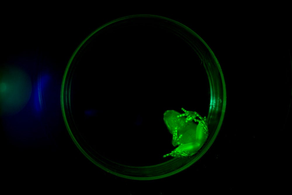
“Wood frog marked with fluorophore”
A.Z. Andis Arietta also won third place in the macro art category. This wood frog was non-invasively marked as a larvae with a fluorophore chelating agent. The fluorophore was absorbed through the tadpole’s skin and bound to calcium stored along the larva’s notochord. During metamorphosis, the fluorophore was imported into the skeletal structure providing a permanent identifying mark for recapture studies. The mark is completely unseen by the naked eye. This is the first application of this type of marking in amphibians and promises to be the only reliable method of marking amphibians that persists across developmental stages.
“Species-level evolutionary relationships and tempo of divergences across global mammals”
Nathan Upham, in the Department of Ecology & Evolutionary Biology and the lab of Walter Jetz, won first place in the illustrations category, first place in the environmental and climate science category, and the competition’s Overall Best Photo award. This is the main figure showing the new Tree of Life of all mammals. It took four years to complete this study. The tree contains 5,911 species of living and recently extinct mammals distributed globally today but extending back to 180 million years of evolutionary history.
“Generative choreography with AI”
Chase Shimmin, in the Department of Physics and the lab of Paul Tipton, won second place in the illustrations category. This is a snapshot showing several frames of a choreographic dance sequence generated by an AI. After learning to statistically represent the space of movements observed in training sessions, the neural network is able to sample and interpolate from a latent posterior distribution to invent its own completely new sequences of movement, or create endless variations of prompt sequences.
“Residential connection: The complex network of first-year college students”
Paul Stillman, in the Department of Marketing and the lab of Ravi Dhar, won third place in the illustrations category. This is a graphical representation of the connection of 102 first-year students living in a residence hall, with each student represented as a point on the perimeter of the circle. Black lines connect the students who indicated knowing one another.
“BigBoi: chicken after dark”
Laurel R. Yohe, in the Department of Geology & Geophysics and the lab of Bhart-Anjan Bhullar, won first place in the category of neuroscience. The orange areas show cell nuclei and the blue areas highlight a protein critical to neural crest development and spinal cord formation.
Media Contact
Jim Shelton: james.shelton@yale.edu, 203-361-8332